

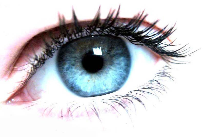
Ever wonder why people have different eye colours?
The answer is in our IRIS because the iris contains pigments that will determine our eye colours.
Do you know that it is possible to have more than just one eye colour?
Wentworth Miller (google him if you don't know him, but i think it is very unlikely because he is just SO famous!) is one good example, but he usually wears contact lens when acting, so that he doesn't appear to be weird.
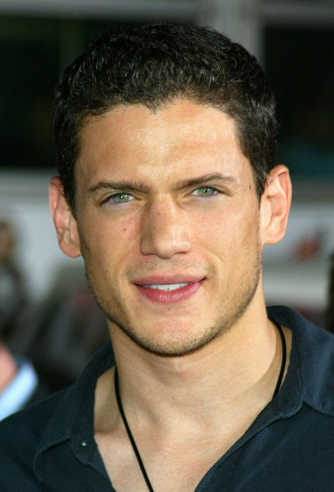
Notice the right eye.

Choroid
- It is pigmented black to prevent internal reflection of light.
- It contains blood vessels which transport oxygen and nutrients to the eyeball and remove metabolic waste products.
Retina
- It is a light-sensitive layer on which images are formed.
- Light-sensitive cells called photoreceptors can be found here.
Photoreceptors
- They are light-sensitive cells which consist of cones and rods.
- They are connected to the nerve-endings from the optic nerve.
Cones
- There are about 7 million cone cells in our eye and they can be found in greatest concentration in the small, central part of the retina called the macula.
- They enable us to see colours in bright light, and do not work well in dim light.
- The three types of cones - red, blue and green cones, contain different pigments which absorb light of different wavelengths.

- All the cones allow us to see a wide variety of colours.
Rods
- There are about 100 million rod cells and they are found in the peripheral retina, away from the macula.
- Compared to cones, they are move sensitive to light and enable us to see in black and white in dim light as they contain a pigment called visual purple.
- When our eye is exposed to bright light, all the visual purple is bleached. Thus, when we enter a dimly-lit room, the visual purple in the rods has to be re-formed before we can see in the dark.
- Vitamin A helps in the formation of visual purple. A lack of vitamin A results in a person suffering from night-blindness.
Macula

- It is a specialized part of the retina.
- It is a patch of densely packed light-sensitive cells which is essential to our central vision and allows us to have a detailed vision.
Optic nerve
- It is a bundle of more than 1 million nerve fibers that exits the back of the eye.
- When the photoreceptors in the retina are stimulated, the optic nerve transmits nerve impulses to the brain where they are interpreted in the primary visual cortex.
Blind spot
- It is the region where the optic nerve leaves the eye.
- It contains neither cones nor rods, hence it is not sensitive to light.
Fovea
- Fovea, or otherwise known as the yellow spot, is the middle part of the macula.
- It is a small yellow depression in the retina and is located directly behind the lens.
- It contains the greatest concentration of cones, but no rods.
- It allows us to have a detailed colour vision in bright light.
 Ciliary body
- It is a thickened region at the front end of the choroid.
- It contains ciliary muscles which control the curvature or thickness of the lens.
- It produces aqueous humor.
Lens
- It is a transparent, circular, biconvex structure, about 10mm in diameter.
- It is elastic, thus it is able to change its shape or thickness so as to refract light onto the retina.
- The change in lens shape allows our eyes to focus on objects at varying distances.
Suspensory ligament
- It is a tissue that attaches the edge of the lens to the cilary body.
Aqueous chamber
- It is the space between the lens and the cornea.
- It is filled with a transparent, watery fluid called aqueous humor.
Aqueous humor
- It is constantly being produced by the ciliary body and drained away.
- It circulates throughout the front of the eye, thus maintaining a constant pressure in the eye.
- It keeps the front of the eyeball firm and refracts light into the pupil. It also nourishes the lens and cornea.
Vitreous chamber
- It is the space behind the lens.
- It is filled with a transparent, jelly-like substance called vitreous humor.
Vitreous humor
- It keeps the eyeball firm and refracts light onto the retina.
Processes which occur that enable us to see: [conclusion]
- Light first passes through the cornea and then through the aqueous humor.
- Next, light passes through the pupil, which is the round opening in the center of the iris.
- Light then penetrates through the lens and passes through the vitreous humor.
- Following this, light reaches the retina where the cones and rods are stimulated to produce a chain of split-second chemical reactions converting light to nerve impulses.
- Finally, nerve impulses are sent through the optic nerve from the retina to the brain where they are interpreted in the primary visual cortex.

Eye movement 
- Each eye has 6 muscles attached to the sclera, thus enabling us to move our eyes and track objects without turning our head.
- The six muscles are: the lateral, medial, superior and inferior rectus muscles, and the superior and inferior oblique muscles.
- These eye muscles work either individually or together, therefore allowing us to shift our field of gaze left, right, up, down and diagonally.
Jia Qian (5) (:
Enjoy (:
xizi
Patients with a colour vision defect caused by disease usually have trouble discriminating blues and yellows. Inherited colour blindness is most common, affects both eyes, and does not worsen over time. This type is found in about 8% of males and 0.4% of females. These colour problems are linked to the X chromosome and are almost always passed from a mother to her son. Colour blindness may be partial (affecting only some colours), or complete (affecting all colours). Complete colour blindness is very rare. Those who are completely colour blind often have other serious eye problems as well.

 Step 2: We now add a digit pattern which is defined by yellow/blue variation only.
  Step 3: Now we add another digit pattern which is defined by red/green variation and is easier to see than the pattern defined by yellow/blue variation.
  Step 4: Finally we add all three components: the random brightness pattern, the yellow/blue pattern, and the red/green pattern. Observers with red/green deficiency will not be able to see the red/green pattern and base their response on the yellow/blue pattern only.
 There is no treatment or cure for colour blindness. Those with mild color deficiencies learn to associate colours with certain objects and are usually able to identify colour as everyone else. However, they are unable to appreciate colour in the same way as those with normal colour vision.
SO, APPRECIATE YOUR EYES!

A RAINBOW (:
♥Love, Nic.
Without our sense of taste, even if the world’s most sumptuous delicacies are placed in front of you, you would not be able to enjoy them. Without our sense of hearing, music would be of no sense to us as we can't enjoy it. Imagine life without our sense of taste, touch and smell and of course, sight.
Blindness is the lack of form and visual light perception. A person who is blind has a high degree of vision loss. Only about 18% of blind people are totally blind - most can distinguish between light and dark.
So how does blindness affect people?
Imagine yourself not being able to see. You have to be extra dependent on your other senses. Simple tasks such as reading, watching television and even eating becomes a chore. Blindness can also cause difficulty with dressing, writing, shopping and going for a walk. The risk of physical and social isolation is greater for people who are blind as it can be difficult to get out and make new friends. Blindness can also be an expensive condition because of the cost of special equipment.
There are many causes as to why people become blind. Accidents and disease are often responsible for blindness, while some people are born blind or partially sighted. Common causes include cataract, macular degeneration, retinitis pigmentosa, diabetic retinopathy and glaucoma. Below, I shall elaborate further on the each of the causes of blindness.
Accidents
One basic cause of blindness can be accidents that injure the eye. Sometimes the eye can be saved and sometimes not with medicines, surgery, etc.. It is very important to do the following to preserve good eyesight:
Do not throw objects in people's faces or at their heads
Be very careful when using sharp objects and point them away from your face
Wear goggles/protective glasses when asked to work with chemicals, fire ...
Cataracts
Cataracts are formed in the lens of the eye, located behind the pupil (as seen from previous posts). As people get older, the lens of the eye gets cloudy. When people have cataracts, it is as if they are looking through a fog. As the cataracts get worse, the fog gets thicker. Although older people more often get cataracts, sometimes kids can have them as well. Your grandparents may get cataracts. Cataracts can be removed by taking out the lens of the eye with surgery and having an artificial lens put in. People can have their vision return to normal after this surgery.
Macular Degeneration
This disease is one of the leading causes of blindness among older people and cannot be cured as of yet. With this disease, the middle of the back of the eye called the macula is diseased.
To understand what a person with macular degeneration sees, make your hands into fists and put your fists right up in front of each eye. Notice that you can see around the fists, but not directly in front of you. People with macular degeneration cannot read or recognise people, but can sometimes move around without as much assistance because the center of the back of the eye is in charge of a person seeing small details that lets them read and recognise people.
Retinitis Pigmentosa
This is a disease that usually begins when a person is young and gets worse as they get older, often leaving them blind as adults.
In the beginning, the person with retinitis pigmentosa may have trouble seeing at night. Later their "field of vision" or the amount they can see in each eye gets less and less. Scientists are not sure exactly how it forms, but at this time there is no cure.
To understand what a person with retinitis pigmentosa may see, roll up a piece of paper and look through the tube you have formed with that paper roll. As the disease gets worse, it is as if the paper roll gets more tightly rolled and the opening gets smaller.
Diabetic Retinopathy
This is caused by the disease diabetes which can strike both children and adults. What happens is that the blood vessels or tubes in the back of the eye break and blood floods into or damages parts of the eye that help a person see. Sometimes surgery or medicines can help a person with diabetic retinopathy and also with the disease diabetes.
Glaucoma
Glaucoma generally affects older people. This happens when fluids in the eye build up and cause too much pressure in the eye, damaging important nerves.
To know what a person feels like with glaucoma, think of how your eyes feel after coming out of a swimming pool with a lot of chlorine in it. (This does not cause glaucoma, but makes the eyes feel as a person does with glaucoma)
If a person who has glaucoma does not take the appropriate medicine for the condition, they can lose their eyesight.
However, Prevention is always better than cure.
Having an eye test at least every two years can help to detect problems that may need treatment before any permanent damage has been done.
It is important to wear protective eyewear in situations where accidental eye damage may occur, for example, at work, when playing sport or doing DIY.
Some conditions, such as glaucoma and cataracts, can be treated to help preserve vision.
♥Love, Nic.
But actually, our eyes do not SEE the objects, they are just the MEDIUM through which we see.
We are able to see because of light, without it, we cannot see.
Our eyes respond to changes in light and enable us to see
- by controlling the amount of light entering the eye, and
- by focusing light reflected from objects onto the retina.
***
In order for us to see clearly, the right amount of light should enter the eye (e.g. less light must enter the eye in bright light).
The size of the pupil determines how much light enters the eye. And the size of pupil is in turn controlled by two sets of involutary muscles in the iris---CIRCULAR MUSCLES AND RADIAL MUSCLES.
Circular muscles are arranged in circles round the pupil, while the radial muscles are arranged radially. They are also called antagonistic muscles because when one set contracts, the other set relaxes and vice versa.

Some of it enters the front of the our eye - the transparent cornea - and is refracted as it meets its curved surface. It then goes through the pupil, and enters the lens, and finally reached the retina.
1) The cornea’s refractive power bends the light rays in such a way that they pass freely through the pupil.
2) The iris works like a shutter in a camera. It has the ability to enlarge and shrink, depending on how much light the environment is sending into the eye.
3) The light rays then strike the lens, which works much like the lens in a camera – shortening and lengthening its width in order to focus light rays properly.
4) The light rays are refracted by the lens and pass through the vitreous humor.
5) Finally, the light rays land and come to a sharp focusing point on the retina, which mainly functions like the film in a camera. It is responsible for capturing all of the light rays, processing them into light impulses through millions of tiny nerve endings, then sending these light impulses through over a million nerve fibers to the optic nerve.
Tidbit: Retina contains light-sensitive cells / photoreceptors, which consist of rods and cones. Cone enable us to see colours in bright light while rods enable us to see in black and white in dim light. The photoreceptors are connected to the nerve-endings from the optic nerve.
6) The optic nerve is like an extension of the brain. It is a bundled cord of more than a million nerve fibers. The nerve impulses travel are transmitted by the nerve fiber to the brain, where they are interpreted as an image.

Now, let's watch a video to learn about how the eyes focus.
Enjoy! (:
The Marine Biological Laboratory (MBL) is an international center for research, education, and tr
 aining in biology, biomedicine, and ecology. It has hosted a long and successful effort to unravel the physics and chemistry of vision.
His student, George Wald, identified many of the molecular components of vision and demonstrated the role that Vitamin A plays in detecting light.
Those with myopia see nearby objects clearly but distant objects appear blurred. With myopia, the eyeball is too long, or the cornea is too steep, so images are focused in the vitreous inside the eye rather than on the retina at the back of the eye. The opposite defect of myopia is hyperopia or "farsightedness" or "long-sightedness" — Normal vision. This is where the cornea is too flat or the eye is too short..

Mainstream ophthalmologists and optometrists most commonly correct myopia throu
 gh the use of corrective lenses, such as glasses or contact lenses. It may also be corrected by refractive surgery, such as LASIK. The corrective lenses have a negative optical power (i.e. are concave) which compensates for the excessive positive diopters of the myopic eye. In some cases, pinhole glasses are used by patients with low-level myopia. These work by reducing the blur circle formed on the retina.

 st worldwide, affecting 28% of school children at the start of their primary education and 70% of those completing university education. Four of 10 adult Chinese people older than 40 years old have myopia. Of greater concern is that a substantial proportion of Singaporeans have high myopia, usually defined as a refraction of -6.0 D or higher. One in 10 adults has high myopia, compared to 1 in 50 in most Western populations. There is also increasing evidence that both prevalence and severity of myopia may be increasing.
More and more children suffer from myopia
GUESSED CAUSES
Racial differences in myopia prevalence between different countries and, in Singapore, between different racial groups, point towards a genetic predisposition to myopia. In a recent study in Singapore, higher rates of myopia were seen in Chinese compared to Indian and Malay school children, despite controlling for education. This points to a strong genetic role.
Besides, compared with other countries, several environment risk factors for myopia, including higher educational attainment, higher socioeconomic status and high amount of near-work activities, are well-developed in children and adult populations.
We are now living in an environment that all you see everyday are nearby. Sitting in front computers. Buildings all around. For small kids, they are spending more time than before on close objects - books, video games, TV, and even computers—the apparent improving standard of living actually gave rise to diminishments in quality of life!
There are new tech to cure myopia, such as laser eye surgery. However, it is of high-risk. True myopia is irreversible. What we need most is prevention. Myopia affects a significant proportion of people of all ages in Singapore. Tackling this problem will necessarily require a ![]() multi-disciplinary approach involving laboratory scientists, clinicians, ophthalmologists and other public healthcare providers.
multi-disciplinary approach involving laboratory scientists, clinicians, ophthalmologists and other public healthcare providers.
Zhujie (27)
Our human eye is the sense organ that gives us our vision. In humans, each eyeball lies in a hollow in the skull known as the orbit and is attached to the skull by rectus muscles. It comprises of three layers, namely the sclera, choroid and retina, all of which have many structures. However, I will first cover on the structures at the front of the eye, which are structures found at the sclera.
Sclera

- It is a tough, white, protective tissue.
- It helps to maintain the shape of the eyeball.
- Muscles which move the eyeball are attached to the sclera.
Cornea
- It is a tough but clear structure.
- It refracts light rays into the eye.
Conjunctiva
Iris
Pupil
Caruncle
Eyelids
- They can be partly closed so as to prevent excessive light from entering the eye and damaging the light-sensitive tissues inside. This is known as squinting.
- Blinking spreads tears over the cornea and conjunctiva and wipes dust particles off the cornea.

Eyelashes
Tear gland